

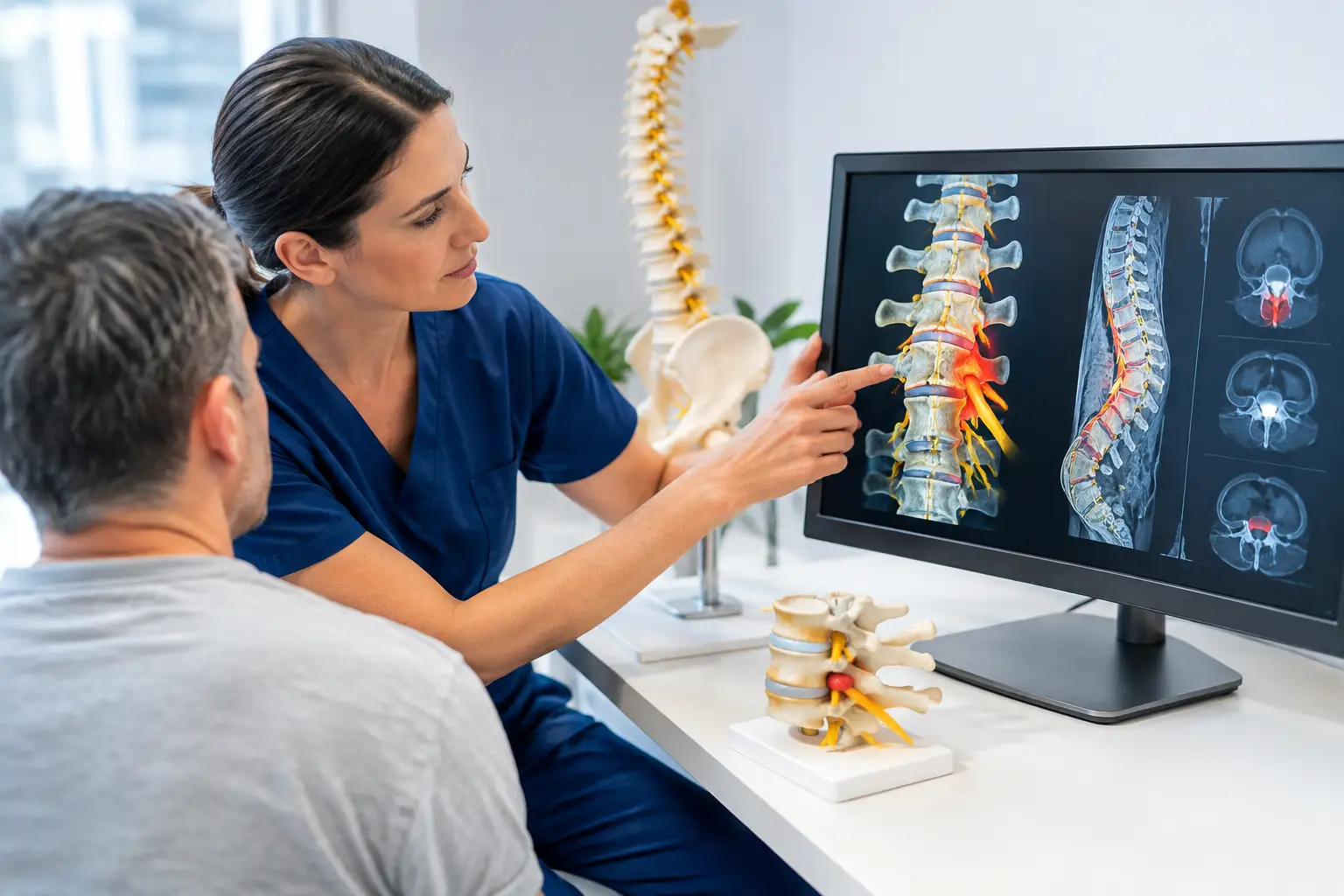
A SEMG 3D back scan can be useful when back pain has become difficult to explain. Perhaps you have had treatment before, your X-ray or MRI has not provided a clear answer, or you know your back feels uneven, stiff or weak but cannot put that feeling into words. A scan can add objective information to a careful physiotherapy assessment – but it is not a diagnosis on its own.
For people living with persistent back pain, sciatica, reduced mobility or recurring flare-ups, the most valuable question is rarely simply, “What does the scan show?” It is, “Does this help explain why I am still struggling to move, walk, sleep or do the things I enjoy?”
What is a SEMG 3D back scan?
SEMG stands for surface electromyography. It is a non-invasive way of recording electrical activity from muscles through sensors placed on the skin. A 3D back scan may combine this muscle activity information with measurements of posture, spinal shape, balance or movement, depending on the equipment being used.
The test does not involve needles, radiation or an injection. Small sensors are placed over selected muscles, usually while you stand in a relaxed position and sometimes while you make simple movements. The assessment is generally comfortable and takes only a short time.
The purpose is to identify patterns rather than to label a single cause of pain. For example, it may show that the muscles on one side of the lower back are working harder at rest, that there is an asymmetry in the way the trunk is held, or that a person is relying on a protective, stiffened movement strategy.
These findings can be relevant, particularly when someone has been in pain for months and has gradually begun moving differently without realising it.
What can a SEMG 3D back scan reveal?
A scan may help a clinician observe muscle activity and posture in a more measurable way. This can be helpful where the body appears to be compensating for pain, previous injury, joint stiffness, reduced confidence in movement or reduced strength.
Common observations may include unequal muscle activity from left to right, excessive resting tension in the back muscles, altered postural loading, or difficulty maintaining a more balanced position. In some cases, a person with leg pain may unconsciously shift their weight away from the affected side. Someone with longstanding lumbar stiffness may hold their lower back rigidly and use the hips or upper back differently to compensate.
That information can support a more personalised rehabilitation plan. Rather than giving everyone with back pain the same sheet of exercises, a physiotherapist can use the findings alongside movement testing, strength assessment, symptom history and day-to-day limitations.
It can also provide a baseline. If you are working on mobility, movement control and confidence after a painful episode, repeating selected measures later may help show whether your physical function is changing alongside your symptoms.
What a SEMG 3D back scan cannot tell you
This distinction matters. A SEMG 3D scan cannot diagnose a disc prolapse, spinal stenosis, arthritis, fracture, infection or cancer. It does not replace an MRI, X-ray, neurological examination or medical assessment where these are needed.
It also cannot prove that a particular muscle reading is the cause of your pain. Muscles often become more guarded after pain begins. Higher activity may be part of the body’s attempt to protect an area, rather than the original problem that needs treating.
Pain is influenced by several factors. These can include joints, discs, nerves, muscle capacity, sleep, stress, previous injuries, general health and how much a person has had to reduce activity. A scan is one useful piece of the clinical picture, not a shortcut to certainty.
This is especially relevant if you have sciatica or nerve-related symptoms. Tingling, numbness, burning pain, weakness or pain travelling into the leg can arise from different mechanisms. Some people need a focused rehabilitation programme, while others may need referral for medical investigation. The right next step depends on the examination, not on one scan result.
Why the assessment around the scan matters
A good assessment starts with your story. When did the symptoms begin? What makes them worse or better? Is walking limited? Does pain wake you at night? Have you noticed weakness, balance changes or altered sensation? What treatment have you already tried, and what happened afterwards?
The physiotherapist then looks at how you move. This may include your walking pattern, spinal movement, hip mobility, leg strength, balance, reflexes and nerve-related signs where appropriate. A SEMG 3D back scan may be used to add measurable information to this process, particularly when posture, muscle guarding or uneven loading appears relevant.
The scan has most value when it changes the clinical conversation. If it confirms an asymmetry that is already visible but does not affect the treatment plan, it is less useful. If it helps explain why a specific movement is difficult, guides the starting point for rehabilitation or makes progress easier to track, it can be worthwhile.
When might a scan be useful for persistent back pain?
It may be worth considering if your back pain keeps returning, you feel stiff or uneven through the trunk, or you have lost confidence in normal movement after an injury or painful episode. It can also be helpful after a period of reduced activity, when deconditioning and protective movement habits may be contributing to ongoing limitation.
Adults over 50 often tell us that the problem is not only pain. It is difficulty getting out of a chair, walking around the supermarket, turning in bed, carrying bags or keeping up with grandchildren. In these situations, the goal is not to chase a perfect-looking scan. It is to understand what is limiting function and create a realistic route back towards independence.
A scan may be less appropriate as a first step if you have symptoms that need prompt medical attention. New loss of bladder or bowel control, numbness around the saddle area, rapidly worsening weakness, fever with severe back pain, unexplained weight loss, significant trauma or a history that raises concern should be assessed urgently by a doctor or emergency service.
How scan findings can guide rehabilitation
If the wider assessment suggests that muscle guarding, poor load tolerance or altered movement control is playing a part, treatment may include hands-on physiotherapy, carefully selected therapeutic exercise and progressive rehabilitation. The precise approach depends on your symptoms, health history and goals.
For one person, the priority may be improving hip movement and walking tolerance. For another, it may be building trunk and leg strength after months of avoiding activity. Where nerve sensitivity is present, the programme may need to progress more gradually, with attention to symptom response rather than forcing painful movements.
Progress is not always linear. A painful back can settle while confidence, strength and endurance take longer to return. Equally, a person may move better before their pain has fully settled. Reviewing both symptoms and function gives a clearer picture than relying on a scan alone.
A clearer starting point for back pain treatment
At NordicFysio, a SEMG 3D back scan is considered within a thorough assessment, not used as a stand-alone answer. For people in Torrevieja, Punta Prima or Ciudad Quesada who remain limited despite previous treatment, that distinction can be reassuring. You deserve an explanation that takes your symptoms, movement and everyday life seriously.
If persistent back pain is stopping you from walking comfortably, sleeping well or enjoying time with the people you care about, a careful assessment can help identify the most sensible next step.